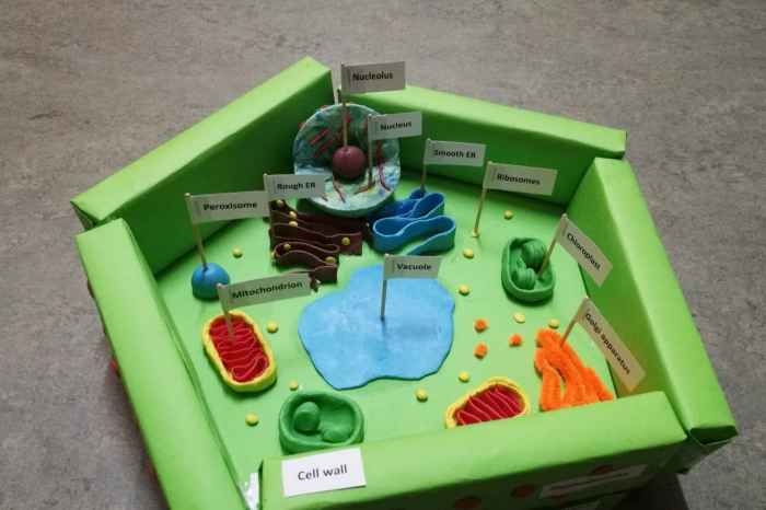
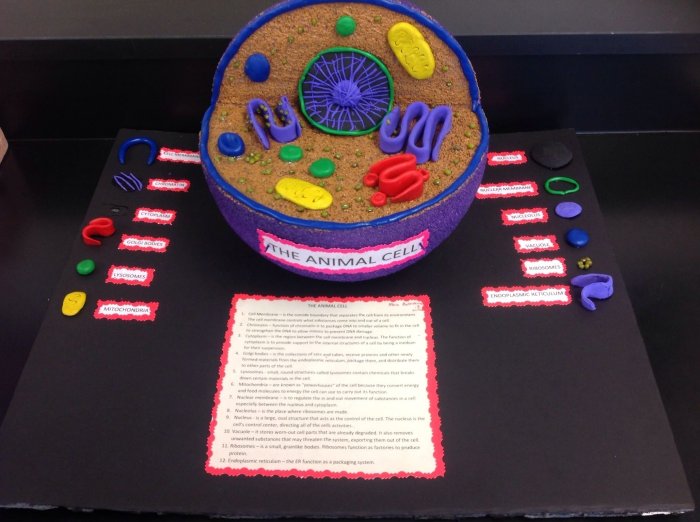
Cell 3D model ideas are a fantastic way to make biology come alive! Imagine diving into the intricate world of cells, exploring their structures and functions in a way that goes beyond static diagrams. With 3D models, you can create interactive and immersive experiences that truly capture the complexity of these tiny building blocks of life.
From visualizing the intricate dance of protein synthesis to understanding the energy-producing powerhouse of mitochondria, 3D models can make complex cellular processes accessible and engaging. They provide a unique opportunity to explore the beauty and wonder of the microscopic world, offering a deeper understanding of the fundamental principles of biology.
Cell Structure and Function
Cells are the fundamental building blocks of all living organisms. They are incredibly complex and organized structures that carry out a wide range of functions essential for life. Understanding the structure and function of cells is crucial for comprehending the intricacies of biology and the mechanisms that drive life processes.
Components of a Cell, Cell 3d model ideas
Every cell, regardless of its type or organism, shares a common set of fundamental components that work together to maintain life. These components, known as organelles, are specialized structures within the cell, each with a specific role to play.
- Nucleus:The control center of the cell, containing the genetic material (DNA) that directs all cellular activities.
- Cytoplasm:The gel-like substance that fills the cell, providing a medium for organelles to function and for chemical reactions to occur.
- Cell Membrane:A selectively permeable barrier that encloses the cell, regulating the passage of substances in and out.
- Ribosomes:Tiny organelles responsible for protein synthesis, the process of building proteins from amino acids.
- Mitochondria:Powerhouses of the cell, responsible for generating energy through cellular respiration.
- Endoplasmic Reticulum (ER):A network of interconnected membranes involved in protein and lipid synthesis, as well as detoxification.
- Golgi Apparatus:A stack of flattened sacs that modify, sort, and package proteins and lipids for secretion or delivery to other organelles.
- Lysosomes:Vesicles containing digestive enzymes that break down cellular waste products and foreign materials.
Role of Cell Components
The intricate interplay between these cellular components is essential for maintaining cell life and function. Each organelle plays a specific role, contributing to the overall harmony of the cell.
- Nucleus:The nucleus safeguards the genetic blueprint of the cell, ensuring the accurate replication and transmission of DNA during cell division. It also directs the synthesis of proteins, the workhorses of the cell.
- Cytoplasm:The cytoplasm provides a stable environment for organelles to function and for chemical reactions to occur. It also serves as a transport medium for molecules within the cell.
- Cell Membrane:The cell membrane acts as a selective barrier, controlling the passage of substances into and out of the cell. This selective permeability is crucial for maintaining the cell’s internal environment and for communication with other cells.
- Ribosomes:Ribosomes are the sites of protein synthesis, a fundamental process that produces the proteins needed for all cellular functions, from structural support to enzymatic activity.
- Mitochondria:Mitochondria are responsible for generating ATP, the energy currency of the cell. They do this through cellular respiration, a process that breaks down glucose to release energy.
- Endoplasmic Reticulum (ER):The ER is involved in the synthesis and transport of proteins and lipids. It also plays a role in detoxification, removing harmful substances from the cell.
- Golgi Apparatus:The Golgi apparatus modifies, sorts, and packages proteins and lipids for secretion or delivery to other organelles. It acts as a distribution center for cellular products.
- Lysosomes:Lysosomes are the cell’s recycling centers, breaking down cellular waste products and foreign materials. They also play a role in programmed cell death (apoptosis).
Cell Membrane: A Vital Regulator
The cell membrane, also known as the plasma membrane, is a vital component that plays a critical role in regulating cell activity. It acts as a selective barrier, controlling the movement of substances in and out of the cell. This selective permeability is essential for maintaining the cell’s internal environment and for communication with other cells.
The cell membrane is composed of a phospholipid bilayer, a double layer of phospholipid molecules. These molecules have a hydrophilic (water-loving) head and a hydrophobic (water-fearing) tail. The hydrophilic heads face the watery environment inside and outside the cell, while the hydrophobic tails face each other in the interior of the membrane.
Embedded within the phospholipid bilayer are various proteins that perform a variety of functions, including:
- Transport proteins:Facilitate the movement of specific molecules across the membrane.
- Receptor proteins:Bind to signaling molecules, triggering specific cellular responses.
- Enzymes:Catalyze chemical reactions that occur at the cell surface.
- Structural proteins:Provide support and shape to the membrane.
The cell membrane’s ability to regulate the passage of substances is essential for maintaining the cell’s internal environment. It allows the cell to take in nutrients, eliminate waste products, and communicate with other cells. This dynamic and complex structure plays a vital role in ensuring the proper functioning of the cell and the organism as a whole.
Cellular Processes: Cell 3d Model Ideas
Cells are not static structures; they are dynamic entities constantly engaged in a variety of processes that sustain life. These processes, ranging from cell division to energy production, are essential for the growth, development, and survival of all living organisms.
Cell Division: The Basis of Growth and Reproduction
Cell division is a fundamental process by which cells reproduce themselves, creating new cells from existing ones. This process is essential for growth, development, and repair in multicellular organisms. There are two main types of cell division:
- Mitosis:A process of cell division that produces two identical daughter cells from a single parent cell. This type of division is responsible for growth, development, and repair in multicellular organisms.
- Meiosis:A specialized type of cell division that produces four daughter cells, each with half the number of chromosomes as the parent cell. This process is essential for sexual reproduction, ensuring the genetic diversity of offspring.
During mitosis, the chromosomes, which contain the cell’s DNA, are duplicated and then separated into two identical sets. The cell then divides, creating two daughter cells, each with a complete set of chromosomes. This process ensures that each new cell receives a full complement of genetic information.
Meiosis is a more complex process that involves two rounds of division. During the first round, homologous chromosomes (pairs of chromosomes with the same genes) exchange genetic material, a process called crossing over. This exchange contributes to genetic diversity. The second round of division separates the sister chromatids (identical copies of each chromosome), resulting in four daughter cells, each with half the number of chromosomes as the parent cell.
Protein Synthesis: Building the Molecules of Life
Proteins are essential for all cellular functions, from structural support to enzymatic activity. The process of protein synthesis, also known as translation, involves the conversion of genetic information encoded in DNA into proteins.
The process begins in the nucleus, where DNA is transcribed into messenger RNA (mRNA). mRNA carries the genetic code from the nucleus to the ribosomes in the cytoplasm. At the ribosomes, the mRNA is translated into a chain of amino acids, which folds into a specific three-dimensional structure to form a protein.
Ribosomes, the sites of protein synthesis, are composed of ribosomal RNA (rRNA) and proteins. They act as the machinery that reads the mRNA code and assembles the amino acid chain.
Cellular Respiration: Generating Energy for Life
Cellular respiration is a complex process that occurs in the mitochondria and is essential for generating energy for all cellular activities. This process involves the breakdown of glucose, a simple sugar, in the presence of oxygen to produce ATP, the energy currency of the cell.
Cellular respiration can be divided into four main stages:
- Glycolysis:Glucose is broken down into pyruvate, producing a small amount of ATP.
- Pyruvate oxidation:Pyruvate is converted into acetyl-CoA, which enters the citric acid cycle.
- Citric acid cycle:Acetyl-CoA is oxidized, producing more ATP and electron carriers (NADH and FADH2).
- Electron transport chain:Electrons from NADH and FADH2 are passed along a series of protein complexes, releasing energy that is used to generate ATP.
Cellular respiration is a vital process that provides the energy needed for all cellular activities, including muscle contraction, nerve impulse transmission, and protein synthesis.
Cell Types and Specialization
Cells are not all the same; they exhibit a remarkable diversity in structure and function. This specialization allows cells to perform specific tasks, contributing to the overall complexity and efficiency of multicellular organisms.
Categorizing Cell Types
Cells can be categorized based on their structure and function. Some common cell types include:
- Muscle cells:Specialized for contraction, enabling movement.
- Nerve cells (neurons):Specialized for transmitting electrical signals, enabling communication within the nervous system.
- Epithelial cells:Form sheets that cover surfaces and line cavities, providing protection, secretion, and absorption.
- Blood cells:Involved in transporting oxygen, fighting infections, and clotting blood.
- Plant cells:Contain chloroplasts for photosynthesis, cell walls for structural support, and vacuoles for storage.
Specialization of Cell Types
The specialization of cell types is achieved through differential gene expression, where different genes are activated or silenced in different cell types. This results in the production of specific proteins that give each cell type its unique structure and function.
Muscle Cells
Muscle cells are elongated and contain specialized proteins, such as actin and myosin, that enable contraction. This allows muscles to generate force and movement.
Nerve Cells (Neurons)
Nerve cells have long, thin extensions called axons that transmit electrical signals over long distances. They also have dendrites that receive signals from other neurons. This allows neurons to communicate with each other and transmit information throughout the nervous system.
Epithelial Cells
Epithelial cells form sheets that cover surfaces and line cavities. They are tightly packed together and have specialized functions depending on their location. For example, epithelial cells in the skin provide protection, while epithelial cells in the stomach secrete digestive enzymes.
Stem Cells: The Source of New Cells
Stem cells are undifferentiated cells that have the potential to develop into different types of specialized cells. They are essential for development and regeneration, replacing damaged or lost cells.
There are two main types of stem cells:
- Embryonic stem cells:Derived from embryos, these cells have the potential to develop into any cell type in the body.
- Adult stem cells:Found in specific tissues, these cells have a more limited potential to differentiate but can still regenerate cells in their respective tissues.
Stem cells hold great promise for regenerative medicine, offering the potential to treat a wide range of diseases and injuries by replacing damaged cells with healthy ones.
3D Model Design Considerations

3D cell models offer a powerful way to visualize and understand the complex structures and processes within cells. Creating realistic and accurate 3D models requires careful consideration of design elements and the use of appropriate software and tools.
Software and Tools for 3D Cell Modeling
A variety of software and tools are available for creating 3D cell models. Some popular options include:
- Blender:A free and open-source 3D modeling software that offers a wide range of features and tools.
- Maya:A professional-grade 3D modeling and animation software used in film, gaming, and other industries.
- ZBrush:A digital sculpting software that allows for detailed and organic modeling.
- 3ds Max:A powerful 3D modeling and rendering software used for creating high-quality visuals.
Key Features and Details

To create a realistic and accurate 3D cell model, it is important to include key features and details that accurately represent the cell’s structure and function. These include:
- Organelles:Each organelle should be modeled with its correct shape, size, and relative position within the cell.
- Cell membrane:The cell membrane should be depicted as a thin, flexible barrier that encloses the cell and regulates the passage of substances.
- Cytoplasm:The cytoplasm should be represented as a gel-like substance that fills the cell and provides a medium for organelles to function.
- Chromosomes:The chromosomes should be modeled as long, thread-like structures that contain the cell’s genetic information.
- Proteins:Proteins should be represented as complex, three-dimensional structures that perform a variety of functions within the cell.
Color, Texture, and Materials
The use of appropriate colors, textures, and materials is essential for enhancing the visual representation of a 3D cell model. Colors should be chosen to accurately reflect the appearance of different organelles and cellular structures. Textures can be used to add realism and detail, while materials can be selected to simulate the properties of different cellular components.
For example, the cell membrane could be represented with a translucent material to highlight its selective permeability. The nucleus could be modeled with a textured surface to represent its complex structure. Mitochondria could be rendered with a reddish-brown color to reflect their energy-producing role.
Applications of 3D Cell Models
3D cell models have numerous applications in education, research, and medicine. They provide a powerful tool for visualizing and understanding the complex structures and processes within cells.
Illustrating Protein Synthesis

A 3D model illustrating the process of protein synthesis could show the following:
- The nucleus, where DNA is transcribed into mRNA.
- The mRNA molecule traveling from the nucleus to the cytoplasm.
- Ribosomes in the cytoplasm, where mRNA is translated into a chain of amino acids.
- The folded protein molecule, representing the final product of protein synthesis.
This model would help students visualize the flow of genetic information from DNA to protein, highlighting the key steps involved in this fundamental process.
Showcasing Cellular Respiration
A 3D model showcasing the interaction between different organelles during cellular respiration could depict:
- Glucose molecules entering the cell through the cell membrane.
- Glycolysis occurring in the cytoplasm, breaking down glucose into pyruvate.
- Pyruvate entering the mitochondria and being converted into acetyl-CoA.
- The citric acid cycle taking place within the mitochondrial matrix, producing ATP and electron carriers.
- The electron transport chain operating on the inner mitochondrial membrane, generating a proton gradient that drives ATP synthesis.
This model would illustrate the complex interplay between different organelles and the flow of energy during cellular respiration.
Demonstrating Plant and Animal Cells
A 3D model demonstrating the structural differences between plant and animal cells could highlight:
- The presence of a cell wall in plant cells, providing structural support.
- The presence of chloroplasts in plant cells, responsible for photosynthesis.
- The larger vacuole in plant cells, used for storage and maintaining cell turgor.
- The absence of these structures in animal cells.
This model would visually emphasize the key differences between these two important cell types.
Outcome Summary
Creating 3D cell models is not just about building virtual structures; it’s about igniting curiosity, sparking creativity, and fostering a deeper appreciation for the incredible world of cells. Whether you’re a student, educator, or simply someone fascinated by the intricacies of life, 3D cell modeling offers a powerful tool for exploration, learning, and sharing the wonders of biology with others.
Quick FAQs
What software can I use to create 3D cell models?
There are many software options available, ranging from free and user-friendly programs like Blender to more advanced professional tools like Maya and 3ds Max. The best choice depends on your skill level and the complexity of the model you want to create.
How can I make my 3D cell model more realistic?
Pay attention to detail! Use accurate dimensions, textures, and colors to create a visually appealing and scientifically accurate representation. You can also add animations to illustrate cellular processes like mitosis or protein synthesis.
What are some real-world applications of 3D cell models?
Beyond education, 3D cell models are used in research, drug discovery, and medical visualization. They can help scientists understand disease processes, test new therapies, and create more effective treatments.